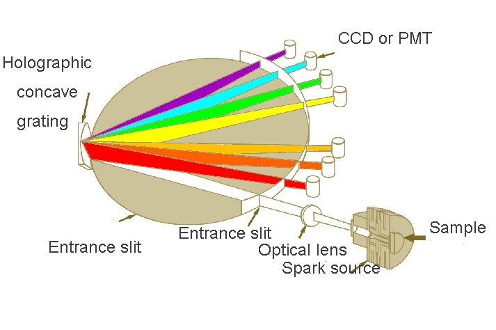
Spectrometer optics
The radiation produced in the radiation source is passed to the entrance slit of the spectrometer optics to be geometrically divided into its constituents (wavelengths). Spectral dispersion takes place in the spectrometer optics. The dispersed radiation is passed behind the exit slit to the radiation receiver and measured values are converted into concentrations using calibration functions- with corresponding corrections.
Radiation guidance to spectrometer optics entrance slit-entrance optics.

In order to obtain calibration curves with minimum SR and identical calibration curves from spectrometer to spectrometer it is necessary to bring the radiation from the entire radiation source homogeneously into the spectrometer optics in order to eliminate changes in intensity caused by displacement of the area of optimum excitation arising from changes in the temperature distribution (geometric and against time) due to third elements and vaporization rate ( but not the concomitant changes in excitation probabilities). This has been known for 60 years but has not been complied with by spectrometer manufactures for the following reasons .First, however, the findings published 60 years ago by Gerlach and Schweitzer.
“If the purpose of the examination is quantitative analysis, it is without question best not to image the spark. Quite apart from the fact that, with imaging, comparison of spectral lines becomes very difficult because of the non-homogeneous intensity distribution along the spark path, the relative intensities in the spectrum, under otherwise identical conditions, are altered by small variations in the imaging process, according to whether imaging is carried out with magnification or reduction, symmetrically or unsymmetrically. Small changes in the sparking process are sufficient for this, and have the same effect as small changes in the imaging process.
The homologous line pairs method is not in principle affected because the emission center of the homologous lines is identical. On the other hand, the optical method of reproducing a spark discharge will not work because a spark line and an arc line are in no way comparable.
Spectra recorded without imaging the spark can be recognized by the complete and symmetrical intensity distribution over the entire height of the line or band”.
Now for the reasons for not complying with the above findings:
Spectrometer radiation sources usually have a non-homogeneous temperature and thus radiation distribution. The temperature gradients may eg. in the case of SDAR, amount to some 1000K/mm .In the radiation sources there are usually regions from which there emanates undesirable background radiation or radiation from multiple-ionised atoms which may raise doubts as to whether analyte lines can be used. Parts of the radiation source are therefore often masked-out with radiation masks or by imaging (or rather non-imaging), which impairs the accuracy of the calibration curves. If masking is not made the same from spectrometer to spectrometer of the same type and for the same analytical task, using the same instrument parameters and samples, the same calibration curves and corrections will not be obtained
The observation zone is the flat surface in the radiation source perpendicular to the optical axis and the surface area. The aperture angle is the solid angle from which the spectrometer can receive radiation while taking measurements. The imaging ratio of the image of the radiation source eg. on the entrance slit or dispersive element, to its natural size.
If, in addition to the discharge parameters, these optical parameters are not held constant from spectrometer to spectrometer, the result will be calibration curve with varying steepness, SR, inter-element effects and LOD.
![]()

Address: 19th Liangtong Road, Mashan Town, Binhu District, Wuxi City, Jiangsu Province, China
Copyright ? 2015 All Rights Reserved
 中文
中文 EN
EN